Scientific Program

Conference Series Ltd invites all the participants across the globe to attend 2nd Annual Conference and Expo on Biomaterials Madrid, Spain.

Day :

  • Track 10: Tissue Engineering and Regenerative Medicine
    Track 14: 3D Printing of Biomaterials
    Track 5: Biomaterials Applications
Speaker

Chair

Elisabeth Engel

University of Catalonia, Spain

Speaker

Co-Chair

Miguel Angel Mateos Timoneda

CIBER of Bioengineering, Biomaterials and Nanomedicine, Spain

Session Introduction

Elisabeth Engel

University of Catalonia, Spain

Title: Tailoring microenvironments for in situ regeneration

Time : 10:45-11:10

Speaker
Biography:

Elisabeth  Engel is an Associate  Professor  at  Technical University of Catalonia since 2010. She received her PhD in 2003 in Bone Metabolism Diseases from a Medical School. She was appointed as PI at the Group of Biomaterials for Regenerative Therapies since September, 2012 at the Institute for Bioengineering of Catalonia. Her research interests include the preparation and design of materials and scaffolds for in vitro and in vivo fundamental studies, and a further focus is the provision of useful tools to assess mechanisms that govern cell behaviour in regenerative medicine.

Abstract:

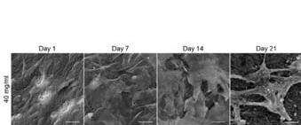
At present, tissue engineering for bone regeneration seeks to obtain scaffolds that mimic the cell microenvironment to recruit stem and progenitor cells to recapitulate the development of target tissues. Herein, we have explored the use of citric acid related to bone nanostructure and mechanical performance, to develop scaffolds resembling the extracellular matrix of developing bone. Elastin-like recombinamers (ELRs) hydrogels were achieved through a one-step chemical crosslinking reaction with citric acid, a molecule currently considered to be essential for the proper performance of bone tissue. We were able to control the architecture and stiffness of citric acid-crosslinked hydrogels while preserving the integrity of adhesion sequences in ELRs. Interestingly, the use of citric acid conferred so-produced hydrogels the ability to nucleate calcium phosphate. In vivo implantation of both mechanically-tailored and non-tailored citric acid-crosslinked hydrogels demonstrated to be able to mineralize the new formed tissue and to integrate into bone in critical size defects in mouse calvaria. Both types of hydrogels showed bone tissue formation by intramembranous ossification. The non-mechanically tailored scaffold showed higher cellular activity (in terms of osteoblasts and osteoclasts presence) related to a lower density of the matrices that allowed higher cell penetration.


Recent Publications:

1. Sánchez Ferrero A, Mata Á, Mateos Timoneda MA, Rodríguez Cabello JC, Alonso M, Planell J, Engel E (2015) Development of tailored and self-mineralizing citric acid crosslinked hydrogels for in situ bone regeneration. Biomaterials 68:42-53
2. Mendes AC, Smith KH, Tejeda-Montes E, Engel, E, Reis RL, Azevedo HS, Mata A (2013) Co-assembled and microfabricated bioactive membranes. Advanced Functional Materials 23(4): 430-438
3. Levato R, Planell JA, Mateos-Timoneda MA, Engel E (2015) Role of ECM/peptide coatings on SDF-1α triggered mesenchymal stromal cell migration from microcarriers for cell therapy. Acta Biomater. 18:59-67
4. Oliveira Hugo, Catros Sylvain, Boiziau Claudine, Siadous Robin, Marti‐Munoz Joan, Bareille Reine, Rey Sylvie, Castano Oscar, Planell Josep, Amédée Joëlle, Engel Elisabeth (2016) The proangiogenic potential of a novel calcium releasing biomaterial: Impact on cell recruitment Acta Biomaterialia 29:435‐445
5. Álvarez  Z,   Castaño   O,   Castells   AA, Mateos-Timoneda MA, Planell JA, Engel E, Alcántara S (2014) Neurogenesis and vascularization of the damaged brain using a lactate-releasing biomimetic scaffold. Biomaterials 3517:4769-4781.

Avelina Sotres Vega

National Institute of Respiratory Diseases, Mexico

Title: Biomaterials as a tool for teaching and learning training programs on surgery

Time : 11:10-11:35

Speaker
Biography:

Avelina Sotres Vega has her expertise in teaching and learning programs on surgery using preserved biomaterials either by cryopreservation or lyophilization as well as cryopreserved tracheal grafts in experimental models of long segment replacement mainly. 

Abstract:

Statement of the Problem: Developing surgical skills is essential in the training of all surgical specialties. However ethical, legal and economic issues have limited surgical training. As surgical educators, we are always trying to identify new ways to provide skills training. We have developed training programs to teach surgical skills to junior surgeons from human and veterinary medicine based on laboratory animals using preserved tissues and organs as surgical training biomaterials. The goal is to help the trainees acquire the abilities and dexterity necessary to perform surgery on patients.

Methodology: We created a small bank of cryopreserved tracheas and stomachs harvested from Wistar rats as well as cryopreserved tracheas and lyophilized esophagus that were harvested from dogs. All animals were previously used in research studies. Stomachs, tracheas and esophagus were cleansed with saline solution, after this, tracheas and esophagus were mounted on polypropylene tubes. In the stomachs, the pyloric antrum was tied with silk 1-0, stomachs were filled with hyaluronic acid solution and the distal esophagus was tied too. Tracheas and esophagus were trimmed in segments of 5 cm. Cryopreservation of tracheas and stomachs was made with a controlled-rate freezer (-1ºC/min) and stored at -30ºC for 30 to 60 days. All the esophagus were lyophilized at -55ºC and 10 mBar of vacuum pressure during 24 hours and sterilized with low temperature hydrogen peroxide gas plasma process. On the day of the surgical skills practice, the cryopreserved organs were thawed at room temperature and all the esophagus were rehydrated with saline solution at 4ºC. Each preserved organ was used to perform end-to-end anastomosis with 4-0 running polypropilene or single stitches. Preserved organs are inexpensive, practical, portable bench models and have high-fidelity that improve the tactile perception and facilitate surgical skills learning by improving how trainees handle tissue and surgical instruments.
 

Recent Publications:
 

1. Sotres-Vega A, Villalba-Caloca J, Azrad-Daniel S, García-Montes JA, Torre-Jaime JL, Guadarrama-Sánchez I, Pérez-Covarrubias D, Santibañez-Salgado JA (2016). Surgical Skills Training with Cryopreserved Rat Stomachs. Journal Veterinary Medical Education 43(4):420-426.
2. Giraldo-Gomez DM, Leon-Mancilla B, Del Prado-Audelo ML, Sotres-Vega A, Villalba-Caloca J, Garciadiego-Cazares D, Piña-Barba MC (2016). Trypsin as enhancement in cyclical tracheal decellularization: Morphological and biophysical characterization. Materials Science & Engineering. C, Materials for Biological Applications (2016) 59:930-7. doi: 10.1016/j.msec.2015.10.094.
3. J. Pineda-Gutiérrez, A. Sotres-Vega, J. Villalba-Caloca, M. Alonso-Gómez, S. Aja-Guardiola, J. García-Jarquín, S. Martínez-Fonseca, J. Salas-Hernández (2016). Preservation of cardiopulmonary blocks: A real and interactive biomaterial for teaching and learning lung plethysmography and mechanical ventilation. International Journal of Engineering Research & Science (IJOER) 2(4):162-166.
4. Olmos-Zúñiga JR, Jasso-Victoria R, Díaz-Martínez NE, Gaxiola-Gaxiola MO, Sotres-Vega A, Heras-Romero Y, Baltazares-Lipp M, Baltazares-Lipp ME, Santillán-Doherty P, Hernández-Jiménez C (2016). Lyophilized allografts without pre-treatment with glutaraldehyde are more suitable than cryopreserved allografts for pulmonary artery reconstruction. The Brazilian of Medical and Biological Research 49(2):e5001. doi: 10.1590/1414-431X20155001.
5. Sotres-Vega A, Osorio-Necoechea ME, Salas-Galindo G, González-Ramón SC, Guadarrama-Sánchez I, Villalba-Caloca J, Santibáñez-Salgado JA (2013). Bench surgical training with lyophilized esophageal segments. Acta Cirurgica Brasileira 28(8):619-23.


Miguel Angel Mateos Timoneda

CIBER of Bioengineering, Biomaterials and Nanomedicine, Spain

Title: Design of a Composite Bioink for Bioprinting Applications

Time : 11:35-12:00

Speaker
Biography:

M A Mateos-Timoneda is an expert in the field of Biomaterials and Scaffolds for Tissue Engineering. He holds a PhD in Supramolecular Chemistry from the University of Twente (Enschede, The Netherlands). Since 2007, he is a Senior Researcher at CIBER en Bioingeniería, Biomateriales y Nanomedicina (CIBER-BBN) and in the Biomaterials for Regenerative Therapies Group at the Institute for Bioengineering of Catalonia (IBEC) in Barcelona (Spain). His main interests are the study of cell delivery using biodegradable microcarriers and 3D printing and bioprinting.

Abstract:

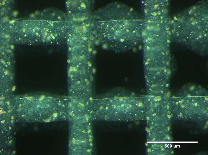
3D bioprinting is an expanding field that allows the fabrication of customized tissue engineered scaffolds with encapsulated cells. Designing a biomaterial fit for 3D printing and cell encapsulation (bioink) is a complex task due to the high number of requirements that need to be accomplished. Bioinks are usually based on combinations of different hydrogels due to their encapsulation capacities, but other kinds of materials can be added in order to improve certain characteristics of the final scaffold. A novel composite bioink that includes alginate as a printable hydrogel and calcium-releasing particles as a vascularization promoter is optimized and studied. Rheometry studies show that the addition of calcium-releasing particles to alginate increases its viscosity, but does not alter its shear thinning properties; therefore maintaining the printability of the material. Solid scaffolds with theoretically high nutrient diffusion rates (filament diameter ≈ 200 µm) are printed using this novel bioink and a novel cross-linking method. A bioprinting procedure that includes encapsulation of cells is optimized and tried out successfully with different bioinks, obtaining good survival rates. The addition of calcium releasing-particles improves cell survival after the bioprinting process as well as during the first culture days. Moreover, the interaction between calcium-releasing particles and alginate is proven to be adequate for bioprinting and could be an interesting line of research for bone regeneration and tissue vascularization applications.
 

Recent Publications:


1. Levato R, Visser J, Planell JA, Engel E, Malda J, Mateos‐Timoneda MA (2014) Biofabrication of tissue constructs by 3D bioprinting of cell‐laden microcarriers. Biofabrication 6:035020.
2. Puñet X, Machauffé R, Giannotti MI, Rodríguez‐Cabello JC, Sanz F, Engel E, Mateos‐Timoneda MA, Planell JA (2013) Enhanced cell‐ material interactions through the biofunctionalization of polymeric surfaces with engineered peptides. Biomacromolecules 14:2690.
3. Levato R, Planell JA, Mateos‐Timoneda MA, Engel E (2015) Role of ECM/peptide coatings on SDF‐1a triggered mesenchymal stromal cell migration from microcarriers for cell therapy. Acta Biomater 18:59.
4. Baelo A, Levato R, Julián E, Crespo A, Astola J, Gavaldà J, Engel E, Mateos‐Timoneda MA, Torrents E (2015) Disassembling bacterial extracellular matrix with DNase‐coated nanoparticles to enhance antibiotic delivery in biofilm infections. J Control Release 209:150.
5. Levato R, Mateos‐Timoneda MA, Planell JA (2012) Preparation of biodegradable polylactide microparticles via a biocompatible procedure. Macromol Biosci 12:557. 

Speaker
Biography:

Sybille Facca, MD, PhD, has her expertise in Orthopedic, Hand and Nerve Surgery as an Orthopedic Surgeon at Strasbourg Hospital University since 2007. She was the first person focusing her research on bone and cartilage regeneration and drug delivery systems of bone cements or nanofibers membranes and osteointegration of orthopaedic implants. Now, she is also focusing her research on tubes for nerve regeneration, microsurgery simulation or microanastomosis mechanical properties and new design of wrist arthroplasty, in a biomechanical laboratory of Strasbourg University.

Abstract:

During the last ten years, tissue engineering has merged with regenerative nanomedicine by combination, not only of new biomaterials but also of stem cell technology and growth factors. The goal of this work was to use bone and cartilage engineering as a model, in order to improve and to develop active and living nanostructured implants. We were interested in the development of biomaterials (natural or synthetic), tridimensional (3D), transplantable for bone and cartilage diseases treatments, that are able to induce more cellular differentiation and improved tissue regeneration. We have developed 3 types of nanostructured implants, (i) titanium implants coated with hydroxyapatite and carbon nanotubes in order to improve osteoformation and osteoinduction around arthroplasty implants; (ii) active capsules functionalized by growth factors and stems cells for bone induction (in vitro/in vivo) after a bone defect; (iii) electrospun nanofibrous membranes functionalized by growth factors and (Osteoblasts/Chondrocytes) for bone and cartilage regeneration, in vitro and in vivo.
Recent Publications:
 

1. Facca S, Lahiri D, Fioretti F, Messadeq N, Mainard D, Benkirane-Jessel N, Agarwal A (2011) In vivo osseointegration of nano-designed composite coatings on titanium implants. ACS Nano 5:4790-4799.
2. Facca S, Lahiri D (2012) Nanoreinforcement of hydroxyapatite coatings on titanium for osseointegration of orthopaedic implants. Comput Methods Biomech Biomed Engin. 15 Suppl 1:10-11.
3. Eap S, Keller L, Ferrand A, Schiavi J, Lahiri D, Lemoine S, Facca S, Fioretti F, Mainard D, Agrawal A, Benkirane-Jessel N (2014) Nanomechanical properties of active nanofibrous implants after in vivo bone regeneration. Nano Life 04(01) DOI: 10.1142/S1793984414500019.
4. Vaiss L, Ichihara S, Ramirez DG, Hendriks S, Liverneaux P, Facca S (2015) A comparative study about ionizing radiation emitted during radiological "skyline" view of the wrist in pronation versus supination. Eur J Orthop Surg Traumatol 25:309-311.
5. Perruisseau-Carrier A, Bahlouli N, Po C, P Vernet, Facca S , Liverneaux P (2017) Analysis of the modifications of MRI signal of the brachial plexus of rats: Comparative study before and after freezing/thawing. Ann Chir Plast Esthet pii: S0294-1260 (16) 30214-X. 


Speaker
Biography:

José Manuel Baena, MSc is a Research Associate. He is the Founder of BRECA Health Care, pioneer in 3D printed custom made implants for orthopedic surgery, and REGEMAT 3D, the first Spanish bioprinting company. He is an expert in innovation, business development and internationalization. He is a Lecturer in some business schools, and  is passionate  about Biomedicine and Technology. In his free time, he also works as a Researcher at the Biopathology and Regenerative Medicine Institute (IBIMER).

Abstract:

Statement of the Problem: The 3D bioprinting of tissues and organs represents a major breakthrough in regenerative medicine and tissue engineering. Cartilage and bone regeneration provides an alternative in the treatment of diseases such as degenerative osteoarthritis, injuries of articular cartilage, osteonecrosis and bone fractures, among others. The purpose of this study is to describe the design, development and preparation of a bioink with hyaluronic acid (HA) to manufacture cartilage and bone by 3D bioprinting.

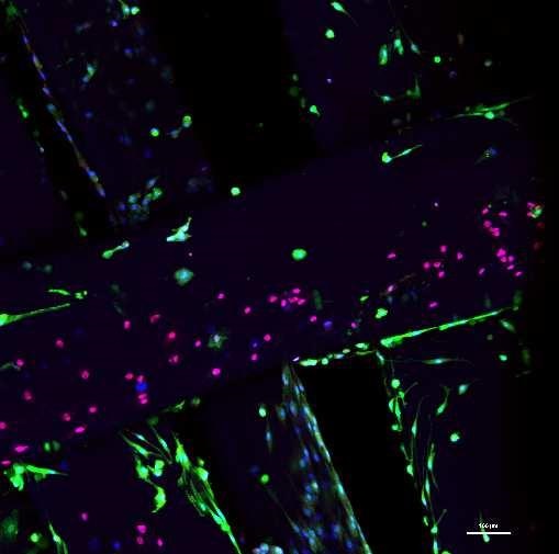
Methodology & Theoretical Orientation: For the formulation of bioinks, two hyaluronic acids were studied: high molecular weight sodium hyaluronate (bioinkA) and low molecular weight sodium hyaluronate (bioinkB), both of intra-articular grade. The HA was combined with alginate and human chondrocytes. The biopaper studied was the polylactic acid (PLA). Cell viability was studied for each bioink.

Findings: The results obtained showed that the HA concentration before and after the bioprinting process did not affect chondrocyte viability. Additionally, cells remained in proliferation after 5 weeks. The rheological properties of each bioink showed mild differences between bioinkA and bioinkB.

Conclusion & Significance: Considering the mild differences in rheological properties between the two experimental bioinks, it may be concluded that both formulations can be used for cartilage and bone bioprinting.
 

Recent Publications:
 

1. Gálvez-Martín P, Hmadcha A, Soria B, Calpena-Campmany AC, Clares-Naveros B (2014) Study of the stability of packaging and storage conditions of human mesenchymal stem cell for intra-arterial clinical application in patient with critical limb ischemia. European Journal of Pharmaceutics and Biopharmaceutics 86:459-468.
2. Gálvez P, Clares B, Bermejo M, Hmadcha A, Soria B (2014) Standard requirement of a microbiological quality control program for the manufacture of human mesenchymal stem cells for clinical use. Stem Cells and Development 23:1074-1083.
3. Gálvez P, Martín MJ, Calpena AC, Tamayo JA, Ruiz MA, Clares B (2014) Enhancing effect of glucose microspheres in the viability of human mesenchymal stem cell suspensions for clinical administration. Pharmaceutical Research 31:3515-3528.
4. Martín MJ, Calpena AC, Fernández F, Mallandrich M, Gálvez P, Clares B (2015) Development of alginate microspheres as nystatin carriers for oral mucosa drug delivery. Carbohydrate Polymers 117:140-149.
5. Llavero F, Urzelai B, Osinalde N, Gálvez P, Lacerda HM, Parada LA, Zugaza JL (2015) Guanine nucleotide exchange factor αPIX leads to activation of the Rac 1GTPase/Glycogen phosphorylase pathway in interleukin (IL)-2-stimulated T cells. Journal of Biological Chemistry 290:9171-9182.

Break: Lunch Break: 12:50-13:50@Zamora

Deniz Yucel

Acıbadem University, Turkey

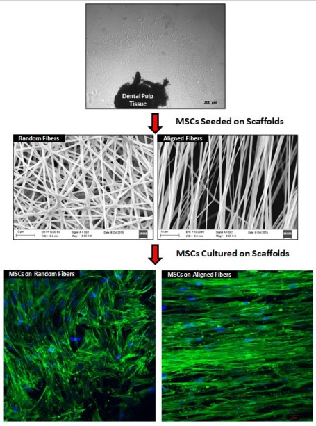
Title: The effect of scaffold topography on behavior of dental pulp stem cells

Time : 13:50-14:15

Speaker
Biography:

Deniz Yucel has expertise in the field of Biomaterials and Tissue Engineering, 2D and 3D polymeric material design, stem cells (mesenchymal stem cells from various sources and neural stem cells), construction of biosensors, and enzyme/protein immobilization on polymeric materials. She received Best PhD Thesis Award in 2010 from Middle East Technical University for her PhD thesis titled, “Stem cell based nerve tissue engineering on guided constructs”. During her PhD, she worked on mesenchymal stem cells and microfluidic systems for one year at Massachusetts General Hospital (MGH) - Harvard Medical School and at Tufts University. She is currently working on stem cells, studying their behavior on different scaffolds, and mainly on tissue engineering applications for various tissues like nerve, bone, tendon, and blood vessel.

Abstract:

Statement of the Problem: Tissue engineering aims to restore the damaged tissues or organs that are incapable of functioning properly. It involves scaffolds seeded with preferably the patient’s own cells like mesenchymal stem cells (MSCs). For guided tissues,like nerve and bone, incorporation of guidance platforms into scaffold designs are known to enhance the regeneration environment. The aligned topography creates a permissive milieu for cell attachment, growth, cytoskeletal organization, guidance and even differentiation. The purpose of this study was to investigate the effect of scaffold topography on behavior of dental pulp stem cells, such as their attachment, proliferation and orientation.
 
Methodology: Random and aligned fibrous mats of polymer blend were fabricated by electrospinning. Human MSCs were isolated from dental pulp tissue. MSCs were seeded and cultured on biodegradable fibrous mats. Proliferation of cells on electrospun mats was studied using MTS. The cytoskeletal and nuclear orientation of the cells on scaffolds were investigated by confocal microscopy after FITC-Phallodin and DAPI staining for cytoskeleton and nucleus.
 
Findings: Random and aligned electrospun fibers without beads were obtained under optimized conditions. MTS results revealed that MSCs were able to grow and increase in number on both random and aligned fibers. Confocal microscopy results demonstrated that MSCs responded to the topography of scaffolds. MSCs on aligned electrospun mats were well oriented along the axis of the fiber while the cells on the randomly organized fibers appeared to spread randomly in every direction.
 
Conclusion & Significance: In the present study, guided tissue engineering approach with MSCs aligned on the highly oriented mats showed that it could serve as a guiding substrate for structural and functional regeneration for oriented tissue injuries.
 

Cristina Plamadeala

Johannes Kepler University Linz, Austria

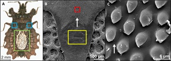
Title: Bio-inspired microstructures for directional liquid transport

Time : 14:15-14:40

Speaker
Biography:

Cristina Plamadeala has a Bachelor’s and Master’s degrees in Biophysics and Medical Physics from the Alexandru Ioan Cuza University, Iasi, Romania. Currently, she is enrolled in the PhD program of Johannes Kepler University Linz, Linz, Austria, under the supervision of a Dr. Johannes Heitz and Dr. Werner Baumgartner. Her scientific work is done in the framework of the European FET-OPEN project 665337 titled, "Laser-induced nanostructures as biomimetic model of fluid transport in the integument of animals (LiNaBioFluid)". The main focus of her work is to create laser-induced microstructures for potential bio-medical applications in the fields of fluid transportation and tissue engineering.

Abstract:

The interdisciplinary field of biomimetics has been very successful in solving engineering problems by searching for solutions in nature. Through the process of evolution, many living organisms developed different structural and chemical material properties that assured the continuation of a certain species. Technological challenges dealing with wetting and liquid collection and transportation also found solutions in nature. Our main focus is on the directional transport of liquids and as a role-model for this application, we used the flat bugs (Dysodius lunatus). Here, we present arrays of microstructures produced by two-photon polymerization technique that mimic the micro-ornamentation from the bugs’ cuticle. A good directionality of liquid transport was achieved, directly controlled by the direction of the pointed microstructures at the surface. These results could therefore be interesting for applications in friction and wear reduction.
 

  • Track 1: Advanced Biomaterials
    Track 7: Biomaterials and Nanotechnology
Speaker

Chair

Monica Lopez Fanarraga

UNIVERSITY OF CANTABRIA-IDIVAL, Spain

Speaker

Co-Chair

Isabel Rodriguez

IMDEA-Nanoscience, Spain

Session Introduction

Mónica López Fanarraga

UNIVERSITY OF CANTABRIA-IDIVAL, Spain

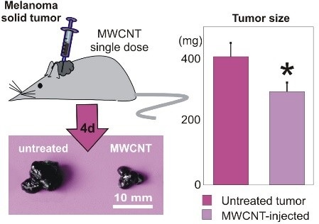
Title: Bio-degradable carbon nanotubes display intrinsic anti-tumoral effects

Time : 14:40-15:05

Speaker
Biography:

Monica L Fanarraga obtained Bachelor’s in Vet. Med. from the University of Zaragoza (Spain), PhD by the University of Glasgow (UK) and Dr. Med. by the University of Cantabria (Spain). Currently, she directs the group of Nanomedicine at the IDIVAL Institute in Santander, Spain. The IDIVAL Nanomedicine group studies the biological response to different nanomaterials focusing in the study of nanomaterials as treatments for cancer, nanotoxicity and nanodelivery.

Abstract:

The complex biosynthetic relationships of nanomaterials with the cellular components, resulting in their chemical nature, surface properties or morphology are often unpredictable. Our research group has demonstrated that Multi-Walled Carbon Nanotubes (MWCNTs) can penetrate inside cells and bind microtubules interfering with the cellular biomechanics. This biomimetic interaction leads to the formation of biosynthetic tubulin polymers displaying an enhanced stability that triggers anti-proliferative, anti-migratory and cytotoxic effects in different types of cancer cells. This antitumor activity is intrinsic to the nature of MWCNTs, complementary and synergetic to that of traditional microtubule-stabilizing anticancer drugs such as Taxol®. A key issue to take into account for the development of new alternatives to traditional drugs based on nanodevices or nanomedicines is the possible long-term effects of these nanomaterials, the tissue accumulation and the elimination rates, just as for traditional cytotoxic chemotherapies, CNTs can also interfere with the function of healthy cells and produce many unwanted side-effects. Consequently, unless most concerns about the toxicity of these materials disappear, the development of new treatments based on CNTs offer a poor risk-to-benefit ratio in oncology. Our group investigates different surface treatments on MWCNTs to make these nanomaterials more biocompatible and biodegradable. Improving in vivo biodegradability of MWCNTs - some of the most resistant materials discovered - is not trivial. Here we show how to preserve the anticancer properties of these nanomaterials, these treatments should maintain the general morphology of the tubes to retain the biomimetics of these filaments with the microtubules. Furthermore, we show how single dosages of o-MWCNTs produce significant anti-tumoral effects in vivo, in solid malignant melanomas produced by allograft transplantation in murine recipients. We believe these findings have critical implications for the development of new CNT-based nanotherapies to overcome drug resistance in cancer among other applications.


Recent Publications:
 

1. García‐Hevia L et al. (2015) Multiwalled carbon nanotubes interfere with cell biomechanics inhibiting cancer cell migration. Adv. Healthc. Mat. 4:1640‐4.
2. Villegas J et al. (2014) Multiwalled carbon nanotubes hinder brain macrophage function interfering with cell migration and phagocytosis. Adv. Healthc. Mat. 3:424-32.
3. García-Hevia L et al. (2015) Anticancer cytotoxic effects of multiwalled carbon nanotubes. Curr. Pharm. Design. 21:1920-1929.
4. García-Hevia L et al. (2016) Multiwalled carbon nanotubes inhibit tumor progression in a mouse model. Adv. Healthc. Mat. 5:1080-7.
5. García‐Hevia L et al. (2016) Nano‐ZnO leads tubulin macrotube assembly and actin bundling triggering cytoskeletal catastrophe and cell necrosis. Nanoscale. 8:10963‐73.


Speaker
Biography:

Isabel Rodriguez is a Research Professor at IMDEA-Nanoscience. Her research interest is on areas related to the application of micro and nano fabrication technologies on polymeric materials to construct functional surfaces. She currently works on the development of antibacterial surfaces and cell culture platforms for cell biomechanical studies. She is also working on the development of multifunctional surfaces, particularly on those with super-hydrophobic, anti-reflective and self-cleaning properties.

Abstract:

Cells can react to synthetic surfaces with a wide way of responses which depend upon many factors, including chemical composition of the surface and the physical properties of the bulk substrate material, including substrate stiffness, topography feature size and geometry. It is now widely accepted that mechanical stimulus exerted onto cells by topographic cues can set off specific physiological processes that ultimately dictate the cell behaviour and fate. Identifying the specific topographical cues that lead to a specific cell behaviour, that is still an endeavour in biomaterial research for application areas impacting regenerative medicine or tissue engineering. In this sense, there have been numerous approaches to develop materials with fine control of the topographical features using micro and nanofabrication techniques. In our laboratory, we use polymer nanoimprinting to produce with nanoscale precision and high reproducibility, cellular instructive micro and nano topographical environments. We specifically investigate the response of progenitor neural stem cells to dense high aspect ratio polymer pillars on the micro and nano scale studies on cell viability, morphology, cell spreading and migration indicating that high aspect ratio topographies impact dramatically the cytoskeleton remodelling and distribution of the cellular tractions which in turn, gave rise to very distinctive cell behaviour. Nanosurface features inspired on the moth eye topography have also been investigated as bactericidal biocompatible surfaces for bionic implants. This surface has been demonstrated to be an effective bactericidal topography against Gram positive and Gram negative bacteria. At the same time, the surface supported cell growth and did not influence the biological cellular responses.


Recent Publications:

 
1. Viela F, Granados D, Osorio M R and Ayuso A, Rodríguez I (2016) Biomechanical cell regulation on high aspect ratio nanoimprinted pillars. Advanced Functional Materials 26: 5599-5609.
2. Ho AY, Luong-Van E, Lim CT, Natarajan S, Elmouelhi N, Low H Y, Vyakarna M, Cooper K and Rodriguez I (2014) Lotus bioinspired superhydrophobic, self-cleaning surfaces from hierarchically assembled templates. Journal of Polymer Science Part B: Polymer Physics 52: 603-609.
3.  Rodriguez I, Lim C, Natarajan S, Ho YY, Luong van E, Elmouelhi N , Low HY, Vyakarnam M and Cooper K (2013) Shear adhesion strength of gecko-inspired tapes on surfaces with variable roughness. Journal of Adhesion. 89: 921-936.
4. Luong-Van E, Rodriguez I, Low HY, Elmouelhi N, Lowenhaupt B, Natarajan S, Lim CT, Prajapati R, Vyakarnam M and Cooper K (2013) Micro- and nanostructured surface engineering for biomedical applications. Journal of Materials Research 28: 165-174.
5.  Lin L, Chu YS, Thiery JP, Lim CT, Rodriguez I (2013) Microfluidic cell trap array for controlled positioning of single cells on adhesive micropatterns. Lab on Chip 13: 714-721. 


Speaker
Biography:

Anna Cifuentes-Rius is an NHMRC Early Career Fellow at the Future Industries Institute and the ARC Centre of Excellence in Convergent Bio-Nano Science and Technology. She is working in the design of three-dimensional nanoarchitectures of bioresponsive nanoparticles, containing drugs and bioactives for a range of therapeutic targets including cancer. Her interdisciplinary research is largely focused on the understanding of the biointerface of advanced bio and nano-materials for the application in the emerging field of theranostics.

Abstract:

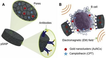
Despite the advances in developing efficient chemotherapy drugs, their efficacy may be diminished by the predisposition of some tumors to drug resistance and non-specific toxicity. Nanomedicine offers the possibility of tackling these key clinical challenges, by designing target delivery platforms for a combination of cancer therapies. To overcome drug resistance, we explore the synergy of hyperthermia with conventional chemotherapy. Due to the higher susceptibility of cancer cells to elevated temperatures compared to healthy cells, hyperthermia stimulates the uptake of anticancer drugs in tumor cells. Thus, nanoparticle (NP)-based delivery systems combining hyperthermia with traditional chemotherapeutics may afford the efficient treatment of highly drug-resistant tumors. Additionally, NP vectorization of therapeutics by actively targeting membrane receptors overexpressed in cancer cells has been recently suggested as a way to ensure selective delivery and improve therapeutic outcomes. Porous silicon (pSi) NPs are (i) biodegradable, (ii) suitable for conjugation with moieties for targeting of a specific cell population, and (iii) exhibit efficient loading of chemotherapy drugs. Here, we utilized these unique characteristics of pSiNPs and loaded them with multiple therapeutics while also immobilizing cell-specific antibodies to achieve active targeting. We have developed antibody functionalized pSi NP loaded with a combination of chemotherapy drug and gold nanoclusters (AuNCs). By selective targeting, these nanocarriers were observed to actively deliver both the chemotherapy drug and AuNCs to human B cells. The accumulation of AuNCs to target cells rendered them more susceptible to the co-delivered chemotherapy drug when an external electromagnetic field in the microwave region was applied. This approach represents a targeted two-stage delivery nanovector that takes advantage of dual therapeutic action in order to enhance cytotoxicity.


Recent Publications:

1.  H Alhmoud, B Delalat, R Elnathan, A Cifuentes-Rius, A Chaix, ML Rogers, JO Durand and NH Voelcker (2015) Title of the abstract. Advanced Functional Materials 25:1137-1145. 


Break: Networking & Refreshment: 15:55-16:10@Zamora
Speaker
Biography:

Michaela Fousova is a PhD student of Materials Science study program at the University of Chemistry and Technology in Prague, Czech Republic. She focuses on metallic biomaterials research and development. The main subject of her professional interest is 3D printing technology applied in the medical sector. She cooperates with industrial companies producing medical implants and also other research centers dealing with metallic additive manufacturing. She already published several papers on the topic of titanium alloy or stainless steel prepared by selective laser melting technology. Recently, she has also got into touch with electron beam melting technology.

Abstract:

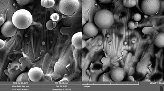
Metallic 3D printing is gaining more and more attention in these days and is gradually becoming a part of industrial production. A lot of metals and alloys can be processed already and there are more than five methods being intensely studied and improved. Application fields of this technology are very wide as almost any shape and design can be achieved. One of the most important fields is biomedicine. With 3D-printed biocompatible metals of desired shape and structure, bone defects can be successfully treated. Although bone structure with gradient porosity can be mimicked, mechanical properties can be adjusted to meet natural properties of the treated bone and osseointegration can be promoted. There are still some drawbacks needing to be solved. Especially in the case of porous structures, there is a problem of unmelted powder particles (being the material input) adhering to the final surface. These particles are harmful for several reasons. Not only they have a negative impact on mechanical performance (particularly fatigue life) and tribological properties, but they might also loosen into the body and set off an inflammatory reaction. Therefore, for biomaterials, surface quality and properties are of a particular importance. Our work focused on titanium alloy Ti6Al4V and its surface morphology and cell compatibility when prepared by 3D printing technology. Although biocompatibility of this broadly used alloy is well known, the interaction with biological environment may be affected by the 3D printing process. For this purpose, samples prepared by two most frequent metallic 3D printing methods – Selective Laser Melting (SLM) and Electron Beam Melting (EBM) were characterized in the as-printed state. Comparison of surface morphology and chemistry has been made. To assess cell compatibility contact in vitro tests were performed.


Recent Publications:
 

1. Fousová M et al. (2017) Promising characteristics of gradient porosity Ti-6Al-4V alloy prepared by SLM process. Journal of the Mechanical Behavior of Biomedical Materials. In press.


Peng-Sheng Wei

National Sun Yat-Sen University, Taiwan

Title: Stages for Pore Formation during fabrication of Porous materials

Time : 16:35-17:00

Speaker
Biography:

Peng-Sheng Wei has received his PhD from Mechanical Engineering Department at University of California, Davis, in 1984. He has been a Professor in the Department of Mechanical and Electro-Mechanical Engineering of National Sun Yat-Sen University, Kaohsiung, Taiwan, since 1989. He has contributed to advancing the understanding of and to the applications of electron and laser beam, plasma and resistance welding through theoretical analyses coupled with verification experiments. He has published more than 80 journal papers, given keynote or invited speeches in international conferences more than 90 times. He is a Fellow of AWS (2007) and a Fellow of ASME (2000). He has also received the Outstanding Research Achievement Awards from both the National Science Council (2004) and NSYSU (1991, 2001 and 2004), the Outstanding Scholar Research Project Winner Award from National Science Council (2008), the Adams Memorial Membership Award from AWS (2008), the Warren F. Savage Memorial Award from AWS (2012) and the William Irrgang Memorial Award from AWS (2014). He has been the Xi-Wan Chair Professor of NSYSU since 2009 and invited Distinguished Professor in the Beijing University of Technology, China, since 2015.

Abstract:

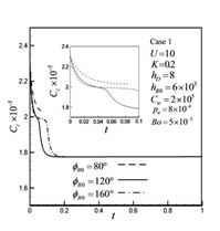
The pore shape in solid as a result of entrapment of a bubble by a solidification front is predicted in this work. Pore formation in solid influence microstructure of materials and contemporary issues of biology, engineering and climate change, etc. This work extends previous models by accounting for realistic mass and momentum transport across a self-consistent shape of the bubble cap. It is found that there exist three stages of solute concentration at the cap or solute gas pressure in the pore during solidification for initial contact angle greater than 90 degrees. Significant drops occur in the early stage and end of a middle stage at which solute concentration at the cap is about that in liquid far from the solidification front and contact angle is near 90 degrees. On the other hand, solute concentration at the cap exhibits two stages in most cases. The predicted pore shape agrees with experimental data. Increases in mass transfer coefficient and solidification rate decrease the pore radius. The predicted pore shape agrees with experimental data.


Recent Publications:
 

1. Wei PS, Chao TC (2016) The effects of drilling parameters on pore size in keyhole mode welding. ASME Journal of Manufacturing Science and Engineering; 138: 021008.
2. Wei PS, Chang CC (2016) Existence of universal phase diagrams for describing general pore shape resulting from an entrapped bubble during solidification. ASME Journal of Heat Transfer; 138: 104503.
3.  Wei PS, Hsiao SY (2016) Effects of mass transfer coefficient on pore shape in solid. International Journal of Heat and Mass Transfer; 103: 931-939.
4.  Wei PS, Hsiao SY (2016) Effects of solute concentration in liquid on pore shape in solid. International Journal of Heat and Mass Transfer; 103: 920-930.


  • Track 2: Polymer Biomaterials
    Track 3: Dental Biomaterials
    Track 4: Properties of Biomaterials
Speaker

Chair

Delair Thierry

University of Lyon, France

Speaker

Co-Chair

Jose Ramon Sarasua

University of the Basque Country, Spain

Session Introduction

Thierry Delair

University of Lyon, France

Title: Polyelectrolyte nano-complexes-Safe and Efficient tools for the delivery of drugs or vaccine

Time : 10:35-11:00

Speaker
Biography:

Thierry Delair received his PhD in Organic Chemistry in 1986 and Post-doctorate at the Stanford Research Institute (California). He has been Professor at University Lyon 1, since November 2008. Previously, he spent 20 years in R&D Department at BioMérieux, a medical diagnostics company. He developed polymeric materials for in vitro diagnostic applications and for vaccine delivery. He has published 135 articles in international peer-reviewed journals (h-index 33), filed 18 patents, and has given 60 oral presentations. His research results encouraged him to establish three companies: Ademtech (magnetic particles), CYTOSIAL BIOmedic (cosmetics SME) and Anabior (vaccines adjuvants).

Abstract:

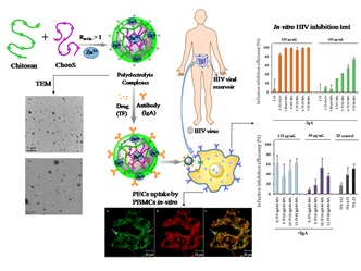
The complexation of polyelectrolytes is very attractive to process polysaccharides into biomaterials, because it is energy efficient, requires no toxic chemical, has a low environmental impact and can be applied to biocompatible polymers such as polysaccharides.We used chitosan, a copolymer of N-acetyl glucosamine and glucosamine obtained from the partial deacetylation of chitin as polycation and a variety of polyanions such as dextran sulphate, hyaluronan, heparin, and chondroitin sulphate. These polysaccharides are generally regarded as safe and some of them can be found in the extracellular matrix of mammals. In this contribution we will present our latest achievement in the control of the elaboration, structure and performances of polyelectrolyte nanocomplexes as drug and vaccine carriers of high potential. In particular, we will address the issues of colloidal stability in physiological media, a major limiting factor in the development of this technology; the nanocomplex loading with drugs or vaccine; the targeting of these nanodelivery systems. The formation of polyelectrolyte complexes is spontaneous at room temperature, i.e. under kinetics control. We will present an alternative approach close to the thermodynamic equilibrium and discuss the potentiality of this particularly innovative synthesis route. Finally, we will present our latest results on the delivery of anti-retroviral drug and the inhibition of the infection by the HIV-1 virus of hPBMCs in vitro.


Recent Publications:
 

1. Wu, D., Delair, T. (2015) Stabilization of chitosan/hyaluronan colloidal polyelectrolyte complexes in physiological conditions, Carbohydrate Polymers 119, 149-158.
2.  Costalat, M., Alcouffe, P., David, L., and Delair, T. (2015) Macro-hydrogels versus nanoparticles by the controlled assembly of polysaccharides, Carbohydrate Polymers 134, 541-546.
3. Costalat, M., David, L., and Delair, T. (2014) Reversible controlled assembly of chitosan and dextran sulfate: A new method for nanoparticle elaboration, Carbohydrate Polymers 102, 717-726.
4.  Polexe, R. C., Terrat, C., Verrier, B., Cuvillier, A., Champier, G., and Delair, T. (2013) Elaboration of targeted nanodelivery systems based on colloidal polyelectrolyte complexes (PEC) of chitosan (CH)-dextran sulphate (DS), European Journal of Nanomedicine 5.
5. Delair, T. (2011) Colloidal polyelectrolyte complexes of chitosan and dextran sulfate towards versatile nanocarriers of bioactive molecules, European Journal of Pharmaceutics and Biopharmaceutics 78, 10-18.


Break: Networking & Refreshment: 11:00-11:15@Zamora
Speaker
Biography:

Jose R Sarasua is Professor of Materials Science at the Department of Mining-Metallurgy Engineering and Materials Science, Faculty of Engineering of Bilbao, the University of the Basque Country (UPV/EHU). He is the Principal Investigator of the ZIBIO group on Science and Engineering of Polymeric Biomaterials and Member of POLYMAT, the Basque Center for Macromolecular Design and Engineering. His research interests are focused on the synthesis, structure and properties of polymeric biomaterials for medical applications.

Abstract:

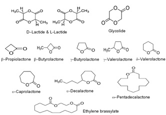
Today’s medicine requires bio-absorbable polymeric biomaterials that present thermoplastic elastomer (TPE) behavior, for their application as medical devices or scaffolds for soft tissue engineering. Among the most investigated polymers used as biomaterials, poly (glycolic acid) (PGA) and poly (lactic acid) (PLA) can be mentioned. These polyesters are however glassy at body temperature and mechanically brittle, so copolymerization with other monomers is a strategy to obtain TPEs with tuned biodegradation rate and mechanical properties. In a search of new polymeric biomaterials with TPE behavior, various lactones and macro lactones, most of them were employed previously by the chemical industry and cosmetics, which draw our attention. Among the cyclic esters, the following ones can be mentioned: β-propiolactone (β-PL), γ-butyrolactone (γ-BL), γ-valerolactone (γ-VL), δ-valerolactone (δ- VL), δ-methyl-ε-caprolactone, decalactones such as γ-decalactone (γ-DL), δ-decalactone (δ-DL) or ε-decalactone (ε-DL) (with rings of 5, 6 or 7 members respectively), ω-pentadecalactone (PDL), hexadecalactone or ethylene brassylate. Moreover, p-dioxanone or trimethylene carbonate may also be of interest. The mentioned substances are monomers that can be synthesized by ring opening polymerization on their own or on the dimmer (lactide and glycolide). In this work TPE copolymers of either high glass transition temperature (Tg) (>20ºC ) or low Tg (between -65 and 0ºC) are synthesized and characterized in terms of molecular parameters, physical, chemical and mechanical properties and biodegradation. In the former case, copolymers of lactide with other co-monomers are proposed in order to reduce the melt temperature and crystallization capability of polylactide. In the latter, alternative copolymers will be introduced for poly (ε-caprolactone). This is because polymers of high Tg present low ductility, brittleness and too high stiffness for soft tissue applications. Those of low Tg, however, though excellent in the combination of mechanical properties for soft tissue engineering and devices, present often too low biodegradation rates.


Recent Publications:


1. Jose R Sarasua, Etxeberria A and Fernández J (2016) Synthesis and properties of ω-​pentadecalactone-​co-​δ-hexalactone copolymers: A biodegradable thermoplastic elastomer as an alternative to poly (ε-​caprolactone). Rsc Advances 6: 3137-3149.
2.  Jose R Sarasua, Fernández J, Amestoy H, Larrañaga-Varga A, Sardon H and Aguirre M (2016) Effect of molecular weight on the physical properties of poly (ethylene brassylate) homopolymers J Mech Behav Biomed Mater 64: 209-219.
3. Jose R Sarasua, Fernández J, Larrañaga A and Etxeberria A (2016) Ethylene brassylate-co-δ-hexalactone biobased polymers for application in the medical field: Synthesis, characterization and cell culture studies. Rsc Advances 6: 22121-22136.
4. Jose R Sarasua, Fernández J, Larrañaga A and Etxeberria A (2014) Tensile behavior and dynamic mechanical analysis of novel poly (lactide/d-valerolactone) statitical copolymers. J. Mech. Behav. Biomed Mater 35: 39-50.
5. Jose R Sarasua and Larrañaga A (2016) Poly (α-hydroxy Acids)-based cell microcarriers. Applied Sciences 6(436): 1-16.


Ruchi Singla

Chandigarh Engineering College, India

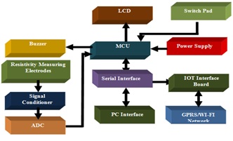
Title: A Novel design of low cost Mastitis level measurement based on electrical resistivity

Time : 11:40-12:05

Speaker
Biography:

Dr. Ruchi Singla is working as Professor and Head of Department in Chandigarh Engineering college, Mohali , India. She has 15 years of work experience and has done PhD in Wireless Communication from Thapar University, Patiala in 2013. She has to her credit around 30 research papers in journals of good repute and filed three patents. Her areas of interest are Antennas and Biosensors.

Abstract:

Mastitis is the major primeval disease of dairy cattle and it leads to inflammation of mammary gland and udder tissue. It is also considered as one of the costliest diseases of dairy animals. Pathogen invades the mammary glands usually caused by bacterial infection of udder tissues. It causes significant harm to the cattleman thereby decreasing the milk production and its quality, which is usually determined by the measurement of somatic cell counts per milliliter of milk. According to the Punjab Dairy Development Board (PDDB), Punjab, India, the average daily milk production in the state is 26.5 million liters a day. Sub clinical mastitis level usually varies from 10% to 50% in cows and 5% to 20% in buffaloes in Punjab region. To minimize the huge economic loss and to provide the cost effective solution for early/preliminary detection of mastitis, the presented work shows an exhaustive survey on cows and buffaloes of Bathlana, Badmajra and Mansa region of Punjab for early detection of mastitis along with a novel design of low cost mastitis detector based on electrical resistivity measurement technique. The presented results show that Buffalo immune system is stronger as compared to cow, due to anatomical structure of mammary glands.
 

Speaker
Biography:

Saurabh Gupta holds Master’s Degree in Oral & Maxillofacial Surgery and has been in surgical practice since 2014. He is also trained in multiple allied surgical disciplines including Implantology and Laser Dentistry. He is involved in cosmetic dentistry including Smile Design (Certified in ClearPath, Botox & fillers). He has published his papers in various national and international journal publications and magazines like “Dentistry Today (US)”, “Access (US)”, etc. He is an editorial board member for many national and international journals (OMICS, Openventio Publishers, Scientia Ricerca, MedCrave, Symbiosis Online Publishing, Bio-Accent, Mathews Open Access Journals, SciFed Dental & Oral Research Journal, etc.). He is also working as a Medical Academic Writer & Research Scientist since 2010 at one of the renowned freelancing sites www.freelancer.com. His research interests are: Dental Implants, Laser Dentistry, Oral and Maxillofacial Surgery, and Dental Pain.

Abstract:

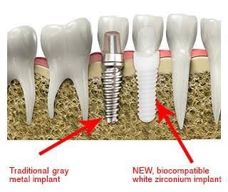
The key aim of this study is to present a comprehensive review of literatures on dental implant materials. The study focuses on titanium implants in a conventional manner and the newly introduced and highly popular zirconia implants. Some of the major areas covered under this study are clinical considerations and material science which includes implant materials as well as the impacts of its physical properties on outcomes of the treatment. Titanium is the gold standard for oral implants fabrication in spite of the sensitivity and despite having unclear clinical relevance. The zirconia implants are highly promising but further clinical studies are required. Also, there is a need of further technical experience and considerations for zirconia implants to lower the mechanical failure incidence.


Recent Publications:


1. S Gupta (2016) A recent updates on zirconia implants: A literature review. Dent Implants Dentures 1:1.
2. Gupta S (2016) Zirconia vs. titanium implants – Deciding factors. J Dent Oral Disord Ther 4(4): 1-2.
3. Saurabh G (2016) Implant stability measure: Critical review of various methods. Dent Implants Dentures 1:110.
4. Gupta S (2015) A review of perimplantitis and lasers. IOSR Journal of Dental and Medical Sciences 14(3) Ver. I:01-04.
5.  Gupta S (2016) Laser-assisted cosmetic dentistry. J Dent Lasers 10:16-8.


Speaker
Biography:

Nour El Houda Kharbech is Young Researcher at the Faculty of Dental Medicine of Monastir in Tunisia. She is a Member of the Tunisian Association of Dental Researches and the Tunisian Red Crescent. She received a Doctoral degree in Dental Medicine from Dental Medicine Faculty in Monastir, Tunisia, recently. Her current field placement is with the Department of Biomaterial in Dental Medicine Faculty at the University of Monastir. She is interested in introduction of nanofillers in the dental field, behavior of PMMA resin under oral conditions, in vitro simulation of oral conditions and removable dental restorations’ problems caused by the material of fabrication.

Abstract:

Statement of the Problem: Despite ceaseless evolutions in the dental practice, PMMA resin has managed to keep a prime position in the panoply of dental materials. Yet, this material is far from being perfect. Concerns are constantly reiterated throughout literature about its lack of mechanical resistance which results in frequent fracture and fatigue failures of dentures. This fracture creates inconvenience to both the patient and the dentist thus it could be considered as a failure of the executed treatment, in addition to further frequently unnoticed consequences regarding the incurred costs to the community of the dentures’ repair. Despite the wide range of solutions suggested to repair the damaged denture and to avoid its further fracture, a consensus seems not to be established yet. Recently, the use of microwave irradiation and nanofillers including silica nanoparticles with PMMA denture-resin has attracted researchers’ attention thus it showed encouraging results in different studies but their efficiency for repairs need to be further investigated.
 
Methodology & Theoretical Orientation: An in vitro study was carried out to investigate the flexural strength, fracture toughness and dimensional stability of 120 repaired PMMA samples after aging in artificial saliva. Autopolymerizing resin was used to repair them following four methods. For the first group, autopolymerizing resin was used alone. In the second, samples were post-treated with microwave irradiations. In the third, the autopolymerizing resin was filled with 2% nanosilica. For the fourth group, the second and the third approaches were combined.
 
Findings: The investigated mechanical properties showed higher mean-values when silica nanoparticles and/or microwave irradiation were used compared to the repair with autopolymerizing resin alone. The dimensional variation rates were under 0.03% for all the groups.
 
Conclusion & Significance: The combination of autopolymerizing resin filled with 2% silica nanoparticles and post polymerization treatment with microwave irradiation showed the highest mechanical properties without affecting the dimensional stability of the repaired samples.
 

Break: Lunch Break:12:55-13:55@Zamora
Poster Presentations:14:00-15:00@Avila
  • Poster Presentations

Session Introduction

Jose Manuel Baena

BRECA Health Care, Spain

Title: Development of scaffolds for Regenerative Medicine
Speaker
Biography:

José Manuel Baena, MSc is a Research Associate. He is the Founder of BRECA Health Care, pioneer in 3D printed custom made implants for orthopedic surgery,
and REGEMAT 3D, the first Spanish bioprinting company. He is an expert in innovation, business development and internationalization. He is a Lecturer in some business schools, and is passionate about Biomedicine and Technology. In his free time, he also works as a Researcher at the Biopathology and Regenerative
Medicine Institute (IBIMER).

Abstract:

Statement of the Problem: Regenerative medicine refers to methods to regenerate or replace human cells, tissues or organs in order to restore or establish normal function. The clinical use of stem cells, genes and tissues constitutes a new range of advanced therapy medicinal products (ATMPs) such as gene therapy medicinal products, somatic cell therapy medicinal products, tissue engineered products and combined advanced therapies’ products. Each of them can be formulated with different types of biomaterials to provide greater cell viability, such as release systems, scaffolds, etc. The purpose of this study is to describe the different products with stem cells and scaffolds that should be considered ATMPs for clinical application, to classify the different types of medicinal products and meet the legal requirements for their marketing authorisation.
 
Methodology & Theoretical Orientation: The aim of this study was to define the main biomaterials used in ATMPs and medical devices, and the regulatory aspects for their clinical application. Thus, we searched the main available databases up to September 2016.
 
Findings: Major advances in advanced therapies focus on the development of matrices made of natural or synthetic origin biomaterials such as collagen, alginate, hyaluronic acid, polyethylene glycol, etc. All of them should be considered medical devices by themselves, but if each scaffold is combined with stem cells, tissues or genes, they will be considered medicinal products.
 
Conclusion & Significance: The ability to combine cells, tissues and genes with biomaterial manufactured structures to develop medicinal products, opens up new prospects in the administration of these ATMPs in the area of regenerative medicine.

Speaker
Biography:

Marco Consumi is a Research Scientist at Department of Biotechnology, Chemistry and Pharmacy in University of Siena, Italy. He has received his PhD in Biomaterials from University of Trento and studied polymers and polymer based materials for controlled release of active substances in pharmaceutical and nutraceutical field. As a Postdoctoral Fellow, he was focused on understanding the correlation between the chemical composition of materials and their biological activity. He has broad expertise in synthesis modification and characterization of polymers (naturals and synthetics) and materials for biomedical applications. Actually, he is involved in 2 EU ITN projects in bacterial infection topic to fundamentally better understand the biology, chemistry and physical properties of biofilms and 2 Toscany Region funded projects on nutraceutical filed.

Abstract:

Monascus fermented red rice (RYR) has been demonstrated to lower cholesterol in blood and sold over-the-counter as an alternative to cholesterol-lowering statin drugs, especially for who stopped statin drugs due to their side effects. The goal of this work is to develop an extended-release formulation, able to maintain the activity effect against the cholesterol, obtaining a constant release of statins present in RYR throughout the staying of the tablets inside the intestine. This study focus on the analysis of different carriers for controlled release systems composed by polysaccharide-based matrices by two different formulations based on K-Carrageenan and Gellan gum (ranging from 10-90% in weight). Samples as cylindrical tablets have been physicochemical characterized by FTIR, DSC, TGA, Rheometer and TOF-SIMS, water uptake, water bond, water diffusion and mesoporosity. The Monacolin K release has been monitored until 48 hours in simulated intestinal fluid SIF. HMG-CoA reductase activity has been measured to determine the formulation influence on statin activities against the Lovastatin activity used as control. The selected formulation enhances the statins release respect to the RYR matrix alone and in addition, the preliminary biological results suggest that the activity of these samples is associated with the inhibition of HMG-CoA reductase. Release tests pointed out that formulations obtained combining polymers in a ratio close to 1 (i.e., 40/60 and 50/50) guaranteed a potentiated release of Lovastatin from RYR inducing also a superior hypocholesterolemizing action both in terms of hepatocytes cholesterol production and inhibitory activity towards 3-hydroxy-3-methylglutaryl-coenzyme A reductase (HMG-CoA). The most effective one, in hypocholesterolemizing activity, in terms of inhibitory activity versus HMG-CoA reductase and hepatocytes cholesterol production, was the formulation obtained combining 40% of K-Carrageenan and 60% of Gellan gum.

Speaker
Biography:

Richard M Hall is a Member of the University of Leeds with an interest in motion preservation devices as well as research in to spinal cord injury and augmentation
procedures such as vertebroplasty. He currently coordinates the LifeLongJoints project and is the Director of Postgraduate Research Studies in Engineering.

Abstract:

Background: We have developed a single method using 3D agarose gels that is suitable to test the biocompatibility of all three types of wear debris (Polyethylene, Ceramic and Metal) simultaneously.
 
Methodology & Theoretical Orientation: Clinically relevant sterile UHMWPE and CoCr wear particles were generated using methodologies described previously. Commercially available nanoscale and micron-sized silicon nitride (SiN) particles (<50 nm and <1 μm, Sigma UK) were sterilized by heat treatment for 4 hours at 180 °C. Agarose-particle suspensions were prepared by
mixing warm 2% (w/v) low-melting-point agarose solution with the particles dispersed by sonication in DMEM culture media. The suspensions were then allowed to set at room temperature for 10 min in 96 well culture plates. Sub-confluent L929 murine fibroblasts were cultured on the prepared gels for up to 6 days in 5% (v/v) CO2 at 37 °C. After incubation, the viability of cells was measured using the ATP-lite assay; the results were expressed as mean±95% confidence limits and the data was analyzed using one-way ANOVA and
Tukey-Kramer post-hoc analysis.
 
Findings: The gels were observed to ensure uniform distribution of particles and migration of cells into the gel. No significant reductions in viability were observed for nanoscale and microscale SiN particles at low doses (0.5 μm3 per cell) and high doses (50 μm per cell) or for UHMWPE wear debris at high doses (100 μm3 per cell). Moreover, the viability was significantly reduced for high doses of CoCr wear debris (50 μm3 per cell) and the positive control, Camptothecin (2 μg.ml-1) at day 6. These results are consistent with the literature and therefore validate our 3D agarose cell culture method.
 
Conclusion & Significance: Biocompatibility of polymer, metal and ceramic wear debris can be tested simultaneously by using 3D particle embedded agarose gels.

Speaker
Biography:

Na Keum Jang received her BS degree in Animal Biotechnology and MS degree at the Department of BIN Fusion Technology, Chonbuk National University, Republic of Korea. Her research interest includes synthesis of polymeric biomaterials for regenerative medicine and sensors applications. Now she is studying microneedle patch for transdermal drug delivery system at Raphas Co. Ltd.

Abstract:

The microneedle-mediated transdermal delivery system has been developed to provide minimal invasive self-administration method with patient friendly manner. Especially, dissolving microneedles, which deliver the target drugs as the drug-loaded microneedle dissolves into the skin, have been spotlighted recently. Droplet-born air blowing (DAB) fabrication method has advantages in stability with precise dose control because DAB provides quick manufacturing process with ambient temperature. The purpose of this study is to show the characteristics of dissolving microneedles, which were manufactured in our mass production system. Microneedle was fabricated by DAB method. The loaded amount of vitamin C and vitamin B3 was analyzed by HPLC/UV system was used to assay the loaded amount within microneedles; and delivered amount of drug into the skin was analyzed using Franz diffusion cell (Logan, FDC-6T). We optimized the DAB process parameters and scaled up. 350 μm and 500 μm length of HA microneedles were fabricated and dried within 10 min without applying any heat. The stability of EGF within HA microneedle was investigated during 2 months at 25, 45°C and was confirmed. The anti-oxidant was stable within HA microneedle during 2 months at 25°C and 45°C. In vitro and ex vivo studies using Franz diffusion cell showed excellent delivery efficiency compared to topical solution. Most of the loaded anti-oxidants was delivered through the skin after 24 hr (98.0±2.0%, n=3). The microneedles dissolution in skin was confirmed, so the drugs within microneedle should be delivered into the intradermal region. Based on the method, we loaded lots of active ingredients with precise dose control, and confirmed the stability of labile drugs such as peptide drugs and antioxidants within microneedles. We are investigating the formulations for biopharmaceutics using this platform technology.

Speaker
Biography:

Patricia Mazón studied Chemistry at the University of Alicante, and pursued her PhD about aminoacids synthesis. She started her adventure in Biomaterials field in 2012 at the University Miguel Hernández. Currently, she is a Professor at the Department of Materials Science, Optic and Electronic Technology and Researcher at the Biomaterials area of Bioengineering Institute.

Abstract:

Alternatives to natural bone grafts are needed in a society that progressively prolonged life expectancy, and it should address the health problems of an aging population. In this context calcium silicophosphate scaffolds are promising candidates. Composition belongs to subsystem Nurse’s A-phase-Silicocarnotite was selected for the ceramic scaffolds that were prepared by the
polymer replication method. An interconnected porous structure with a striking similarity to human cancellous bone tissue was obtained. Response to ceramic scaffolds was evaluated by implantation in New Zealand tibia rabbits in periods of 3 and 6 months. Radiological studies showed correct integration and partial resorption of the scaffold. Histological results presented no evidence for
inflammation or infection at the implantation sites. Colonization process of the scaffold started in the periphery and then penetrated throughout implant porosity. Scaffolds degraded over time and that degradation happened according to the tissue in-growth rate. Histomorphometric analysis gave high BIC values (67.30% ± 1.41) opposite to control samples, where newly formed bone in the cortical defect increased in a smaller amount than in the grafted defects. After six months of implantation SEM studies reveal that the whole ceramic implant surface was covered by a newly formed bone tissue. The new bone layer was composed of Ca-P, mainly with traces of Si due to the gradual diffusion of Si ions from the scaffolds into the newly forming bone, which formed part of the biomaterial’s resorption process. The results indicate that this material provides an optimal microenvironment for the osteogenic differentiation of the undifferentiated osteoblastic precursor cells contained in hematopoietic bone marrow.

Speaker
Biography:

Anna Arvidsson has her expertise in biomaterials and medical device surfaces, with a specific interest related to infection. She has an interdisciplinary background within engineering biology, directed towards biomaterials. During years at Göteborg University with research on surface mediated interactions at the bone/soft tissue implant interface, she has gained experience in surface modifications, surface characterization, and in vitro and in vivo models. Based on this knowledge, she is now creating and exploring new innovations at Dentsply Sirona Implants with purpose to further improve dental implant treatments and tissue regeneration.

Abstract:

Statement of the Problem: Biomaterial associated infections are challenging to treat due to bacterial biofilm formation. Infections around dental implants (so called peri-implantitis) may occur both shortly after implantation as well as after several years. Some risk factors are patient related, such as a prior history of periodontitis, poor oral hygiene, and smoking. However, product properties have also been indicated to influence the risk, progression, and resolution of peri-implantitis. It is hypothesized that anti-biofilm surface modifications may have a counteracting effect in the progress of infection and thereby reducing the risk of infection. Titanium nitride (TiN) coatings are used for dental abutments due to its golden color, but have also been found to accumulate less amounts of plaque. However, there is limited knowledge on possible mode of action. The purpose of this study is to characterize the surface properties of
TiN and to investigate biofilm formation on TiN in comparison with Ti.
 
Methodology: Pre-conditioned specimens were incubated with a co-culture of different oral bacterial species for up to four days. Biofilm formation was evaluated with plate counts, qPCR, live/dead, and crystal violet. Ti and TiN specimens were also characterized using a set of different surface analytical techniques.
 
Findings: After 24 h, plate counts showed a log 2 reduction of bacterial load on TiN compared to Ti, while qPCR failed to show a difference. Live/dead indicated that the biofilm is thinner on TiN than on Ti.
 
Conclusion & Significance: TiN was found to have a certain degree of anti-biofilm properties in vitro. However, any conclusions on clinical significance need data from randomized clinical studies.

Speaker
Biography:

Richard M Hall is a Member of the University of Leeds with an interest in motion preservation devices as well as research in to spinal cord injury and augmentation procedures such as vertebroplasty. He currently coordinates the LifeLongJoints project and is the Director of Postgraduate Research Studies in Engineering.

Abstract:

Adverse biological responses to wear debris generated by total hip replacements (THRs) limit the lifetime of such devices. This has led to the development of biocompatible coatings for prostheses. Silicon nitride (SiN) coatings are highly wear resistant and any resultant wear debris is soluble, reducing the possibility of a chronic inflammatory reaction. SiN wear debris produced from coatings has not been characterized in vivo. The aim of this research is to develop a sensitive method for isolating low volumes of SiN wear debris from periprosthetic tissue. Commercial silicon nitride particles of <50 nm (Sigma Aldrich) were incubated with formalin fixed sheep synovium at a volume of 0.01 mm3/g of tissue (n=3). The tissue was digested with papain (1.56 mg/ml) and proteinase K (1 mg/ml) and samples were subjected to density gradient ultracentrifugation using sodium polytungstate (SPT) to remove protein from the particles. Control tissue samples, to which no particles were added, were also subjected to the procedure. Particles were washed to remove residual SPT and filtered onto 15 nm filters. The filtered particles were imaged by scanning electron microscopy and positively identified by elemental analysis before and after the isolation procedure. To validate whether the isolation method affected particle size or morphology, imaging software (imageJ) was used to determine size distributions and morphological parameters of the particles. A Kolmogorov-Smirnov test was used to statistically analyze the data. The particle size distributions of isolated and nonisolated particles were similar. Morphology in terms of roundness and aspect ratio was unchanged by the procedure. Future work aims to test the method on titanium and cobalt chrome wear debris generated by a pin-on-plate wear simulator. The method will then be applied to isolate and characterize particles from in vivo studies of novel SiN coated prostheses in a rabbit and sheep model.

Speaker
Biography:

Abstract:

The microneedle-mediated transdermal delivery system has been developed to provide minimal invasive self-administration method with patient friendly manner. Especially, dissolving microneedles, which deliver the target drugs as the drug-loaded microneedle dissolves into the skin, have been spotlighted recently. Droplet-born air blowing (DAB) fabrication method has advantages in stability with precise dose control because DAB provides quick manufacturing process with ambient temperature. The purpose of this study is to show the characteristics of dissolving microneedles, which were manufactured in our mass production system. Microneedle was fabricated by DAB method. The loaded amount of vitamin C and vitamin B3 was analyzed by HPLC/UV system was used to assay the loaded amount within microneedles; and delivered amount of drug into the skin was analyzed using Franz diffusion cell (Logan, FDC-6T). We optimized the DAB process parameters and scaled up. 350 μm and 500 μm length of HA microneedles were fabricated and dried within 10 min without applying any heat. The stability of EGF within HA microneedle was investigated during 2 months at 25, 45°C and was confirmed. The anti-oxidant was stable within HA microneedle during 2 months at 25°C and 45°C. In vitro and ex vivo studies using Franz diffusion cell showed excellent delivery efficiency compared to topical solution. Most of the loaded anti-oxidants was delivered through the skin after 24 hr (98.0±2.0%, n=3). The microneedles dissolution in skin was confirmed, so the drugs within microneedle should be delivered into the intradermal region. Based on the method, we loaded lots of active ingredients with precise dose control, and confirmed the stability of labile drugs such as peptide drugs and antioxidants within microneedles. We are investigating the formulations for biopharmaceutics using this platform technology.

Speaker
Biography:

Eloisa Gonzalez-Lavado is a PhD student in the Nanomedicine Group of the University of Cantabria (Spain). I did a chemistry Bachelor’s degree at the University of Extremadura (Spain).I have an european interuniversity master’s degree in theoretical chemistry and computational modelling. Currently I am working with carbon nanotubes and their biomedical aplications, especially in Cancer, studying their biocompatibility, their capability as nanocarriers and their own antitumoral effect.

Abstract:

Our laboratory has focused on the intrinsic anti-proliferative, anti- migratory and cytotoxic effects of carbon nanotubes (CNTs). We have shown how MWCNTs interact with microtubules assembling biosynthetic polymers triggering serious biomechanical cellular defects that lead to cancer cell death. These properties of CNTs produce antitumoral effects in solid melanoma tumors in vivo. The huge surface area of CNTs maximizes their ability to interact with many biological components and different chemicals, constituting their biocorona. Taking into account these surface properties, we aimed to increase these intrinsic antitumoral effects of CNTs functionalizing these nanomaterials with a well-known anti-tumoral drug (5-fluoracil) in vitro in melanoma cells and in vivo in solid malignant melanomas produced by allograft transplantation in murine recipients. We have double-coated CNTs with an internal chemical layer surrounded by a second coat of proteins. The first layer carrying chemicals, either a dye (as a proof-ofconcept) or a drug (5-fluoracil) and the second being a serum protein coating layer, both assembling the biocorona. The protein coating serves for (1) CNTs recognition by cellular receptors, (2) endocytosis, (3) protection of the chemical component attached to the nanotube surface until protein degradation that takes place at the lysosome, and (4) the release of the transported drug during the first 5-9 hours next to the internalization process. CNTs loaded with 5-fluoracil double coated with serum proteins display a significantly enhanced antitumoral effect in vitro and in vivo in mice bearing solid melanoma tumors.

Iordana Neamtu

Petru Poni Institute of Macromolecular Chemistry, Romania

Title: Nanogels preparation for controlled bioactives’ delivery
Speaker
Biography:

Iordana Neamtu is a Senior Scientific Researcher at “Petru Poni” Institute of Macromolecular Chemistry in Iasi - Romania. Her expertise is in synthesis of polymer materials with potential biomedical applications. She has published more than 50 papers in reputed journals and participated in more than 15 Romanian Projects.

Abstract:

Nanogels are cross-linked polymeric particles, which can be considered as hydrogels if they are composed of water soluble/ swellable polymer chains. Their applications for polymer-based bioactives’ delivery systems require biodegradability, controlled particle size with uniform diameter, large surface areas for multivalent bioconjugation and interior network for the incorporation
of different therapeutics, environmental-stimuli responsive capability, dispersibility in biological fluids, sustained release in time of bioactives and facile removal of the devices after the bioactives delivery, etc. This study describes the synthesis and characterization of a stimuli-responsive nanogel performed by crosslinking poly(itaconic anhydride-co-3,9-divinyl-2,4,8,10-tetraoxaspiro [5.5] undecane) with 1,12 dodecandiol. The copolymer with different ratios between the two comonomers is able for network formation,
binding properties, amphilicity, and good oxidative and thermal stability. At the same time the new nanogel structure has high functionality, biocompatibility, temperature and pH responsivity, and is designed to have potential biomedical applications. The chemical structure is explored utilizing common spectroscopic analyses, while the dual pH and temperature sensitivity is evaluated by determining the hydrodynamic radius and zeta potential by dynamic light scattering technique. The analysis of the thermal stability by thermogravimetric analysis supports the new covalent bonds realized by the crosslinking reaction between the copolymer and diol. The acute toxicity of the nanogel is estimated after mice oral administration. Accordingly, analysis of histological evaluation of liver tissues does not reveal substantial pathological modifications. The results propose that the nanogel may be suitable for in vivo
use as bioactives’ delivery system.

Break: Poster Presentations:15:10-16:00@Avila
Speaker
Biography:

Tiago Monteiro is a second year PhD student at the Doctoral Program in Sustainable Chemistry at Faculdade de Ciências e Tecnlogia, Universidade Nova de Lisboa, Portugal. He has previously obtained a MSc degree in Biotechnology (2013) and a BSc degree in Molecular and Cellular Biology (2011) at the same
institution.

Abstract:

Real world application of reductase-based electrochemical biosensing devices is limited by the need of anaerobic working conditions. Molecular oxygen is a main interferent because (a) it can react with many redox mediators in their reduced form, and (b) the reduction of oxygen at the working electrode surface generates an intense background noise that can mask important redox processes occurring between -200 and -800 mV (vs SHE). Standard laboratorial oxygen removal strategies, such as argon purging or vacuum degassing, are incompatible with on-site monitoring. Alternatively, a bi-enzymatic scavenging system that efficiently reduces the soluble oxygen content in small volume samples, and can maintain anaerobic conditions in an open-air environment for extended periods of time, was adapted for biosensing purposes. The scavenging system is composed by glucose oxidase (GOx) and catalase (Cat) in solution and uses the glucose as main substrate, removing oxygen in a two-step cycle. This strategy was successfully employed with a miniaturized reductase-based biosensing tool to monitor nitrite in real samples. We now aim at making the scavenging system an integral part of the biosensors, simplifying operating procedures and reducing costs. Therefore, we have immobilized GOx and Cat on pyrolytic graphite electrodes, using a silica sol-gel matrix, and tested the system’s ability to provide local anaerobic conditions. Several combinations of GOx/Cat in solution and in the immobilized state were prepared and the electrochemical response, in non-deareated solutions containing glucose, was monitored by cyclic voltammetry. Our results showed that although the scavenging system was more effective when in solution, the co-immobilized GOx and Cat were still able to provide a satisfactory anaerobic environment. Additionally, the configurations containing either immobilized GOx or Cat were explored as biosensing tools to monitor glucose and hydrogen peroxide, respectively. The GOx-based bioelectrode responded linearly to glucose concentrations from 1.2 – 7 mM.

Aurica P Chiriac

Petru Poni Institute of Macromolecular Chemistry, Romania

Title: New method for magnetic composite deposition onto a stent surface
Speaker
Biography:

Aurica P Chiriac has completed her PhD in 1994. She has published more than 100 papers in reputed journals and is an Editorial Board Member of some reputed journals. She participated in more than 15 Romanian Projects and 5 European Projects.

Abstract:

The study is devoted to investigating the possibility of using an alternating magnetic field (AMF) for deposition and covering a stent surface and improve its functionalities with a new magnetic composite (MC). MC was prepared in situ during functionalization of poly(maleic anhydride-co-3,9-divinyl-2,4,8,10-tetraoxaspiro [5.5] undecane) copolymer by opening the anhydride ring with
erythritol and introducing magnetic nanoparticles into the polymer matrix. Ten different solvents were used to evidence the dependence between AMF presence, the reaction medium characteristics and the kinetic deposition. Interdependence among the viscosity, density and molar polarization of the solvents and the yield of deposition was registered. The covering of stent with MC is also analyzed by microscopy and the new magnetization values are estimated.

Speaker
Biography:

Abstract:

Statement of the Problem: Biomaterial associated infections are challenging to treat due to bacterial biofilm formation. Infections around dental implants (so called peri-implantitis) may occur both shortly after implantation as well as after several years. Some risk factors are patient related, such as a prior history of periodontitis, poor oral hygiene, and smoking. However, product properties have also been indicated to influence the risk, progression, and resolution of peri-implantitis. It is hypothesized that anti-biofilm surface modifications may have a counteracting effect in the progress of infection and thereby reducing the risk of infection. Titanium nitride (TiN) coatings are used for dental abutments due to its golden color, but have also been found to accumulate less amounts of plaque. However, there is limited knowledge on possible mode of action. The purpose of this study is to characterize the surface properties of
TiN and to investigate biofilm formation on TiN in comparison with Ti.
 
Methodology: Pre-conditioned specimens were incubated with a co-culture of different oral bacterial species for up to four days. Biofilm formation was evaluated with plate counts, qPCR, live/dead, and crystal violet. Ti and TiN specimens were also characterized using a set of different surface analytical techniques.
 
Findings: After 24 h, plate counts showed a log 2 reduction of bacterial load on TiN compared to Ti, while qPCR failed to show a difference. Live/dead indicated that the biofilm is thinner on TiN than on Ti.
 
Conclusion & Significance: TiN was found to have a certain degree of anti-biofilm properties in vitro. However, any conclusions on clinical significance need data from randomized clinical studies.

Speaker
Biography:

Zeynep Orman has graduated from Chemical Engineering department and currently studying Bioceramics for her Master’s studies in Yildiz Technical University. She is experienced in polymers research and has conducted a project about solar cell studies.

Abstract:

The regeneration potential of human bone to repair large bone defects often requires biomaterials, such as those associated with comminuted fractures or bone tumor resection. The need for bone substitutes is rapidly increasing and many researches have been done to improve their performance. Calcium phosphate based bioceramics have been used due to their high bio-compatibility and their successful use of orthopedic and dental applications. Calcium phosphate bioceramics are similar to bone and tooth minerals due to their chemical and crystallographic properties; hence it is particular interest for bone grafting, augmentation in maxillofacial surgery and in orthopedics as a filling material. Many marine structures are composed of calcium carbonate (aragonite or calcite) and can be converted to calcium phosphate materials by chemical exchange. In this study structural and chemical properties of Clinocardium ciliatum based bio ceramic materials (TCP, B-TCP and other phases) were produced by using mechano-chemical (hot-plate) conversion method. At three varying temperature of 450°C, 850°C and 1200°C the materials were transformed to various bioceramic phases. For complete characterization of the bioceramics produced Fourier Transform Infrared Spectroscopy (FTIR), x-ray diffraction (XRD), Brunauer–Emmett–Teller (BET) and differential thermal analysis (TG/DTA) analyses were carried out.

Speaker
Biography:

Loredana E Nita is currently a Senior Researcher at Petru Poni Institute of Macromolecular Chemistry, Romania. She has completed her PhD in Chemistry in 2007 at Petru Poni Institute of Macromolecular Chemistry. She has published more than 100 papers in reputed journals and is a member in Editorial Board of some reputed journals. She participated in more than 15 Romanian Projects and 5 European Projects.

Abstract:

In the last decades, considerable attention has been dedicated to study of interactions between polymer-polymer pair and interpolymer complexes. In this contest, the self-assembling procedure for preparation of functional nanostructure based on linear polyampholyte polypeptide, poly(aspartic acid) (PAS) and a globular protein, bovine serum albumin (BSA), have been studied. The main interest was to identify the formation of an interpenetrated complex between a natural protein and a synthetic polymer in order to design materials suitable for biomedical applications, such as carriers for drug delivery. From the viscometric investigation of PAS/BSA/water ternary systems, it was observed that, for x* value near 0.5, the maximum of intermolecular interactions among the two polymeric partners take place. This statement is sustained by the strong raise of the hydrodynamic radius in the same area of composition. Dielectric spectroscopy data also provide the higher compatibility of PAS with BSA protein molecules and confirm the best conditions for a stable interpolymer complex formation by self-assembling at the PAS/BSA molar ratio x* of 0.542 in aqueous solution. Thus, it is quite important for having desired properties - chemical and biological - the setting and fulfillment of the conditions for achieving the non-covalent forces to make highly functional nanoscale compounds.

Speaker
Biography:

Shghayageh Javadi did her Master’s degree in Biomedical Engineering. Her thesis is about biomaterial coating and focused on best coating method of biomaterials. She has researched on different pretreatment for surface modification of titanium. She has used anodizing process with combination of alkali treatment for surface modification of titanium. The result of her search and experiments showed that the anodized titanium at high voltages may be suitable substrate for biomimetic coating procedure.

Abstract:

Biomimetic coating procedure was used to deposit a bioactive layer of hydroxyapatite (HA) on the surface – modified titanium by anodization process. The anodization was performed on the clean surface of titanium under three different voltages of 80, 100 and 130v using HF as the electrolyte. The morphology, structure and topography of the anodized substrates were evaluated by Scaning Electron Microscopy (SEM) or Field Emission Scanning Electron Microscopy (FE-SEM), X ray Diffraction (XRD) and Atomic Force Microscopy (AFM), respectively. Biomimetic coatings were deposited on the surface of anodized titanium using a twostage procedure by immersion in two concentrated Simulated Body Fluids (SBF) with different concentration of Mg+2 and HCO3-2 ions under physiological conditions. The results showed that an increase in anodization voltage tends to produce a porous surface with circular pores toward a columnar layer of rutile. The surface roughness of anodized surfaces was increased by increasing voltage. Biomimetic coating procedure was caused to form a HA layer which was proved by XRD, FTIR and SEM. The HA layer formed on the anodized titanium surface has a whisker-like morphology. The crystallinity of HA layer was increased by an increase in voltage. The findings indicate that the anodized titanium at high voltages may be suitable substrate for biomimetic coating procedure.

Break: Networking & Refreshment: 16:00-16:15@Zamora
Award & Closing Ceremony: 16:15 onwards@Avila